But most of the people who got the disease were vaccinated for it!

If you follow the social media postings of various anti-vaccination groups, you’ve probably seen them point to contemporary outbreaks of disease, and say something along the lines of:

While the media seem to be pushing the line that the unvaccinated are responsible for bringing these diseases back, when you look at the numbers from [particular outbreak], in fact most of the cases of the disease were in people who received the vaccine! [link to a news article with said statistic] Clearly this vaccine isn’t working if the vaccinated make up most of the notifications.

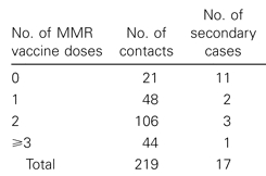
So, is this a valid point? On what basis are vaccines even considered to be effective, and how does that determination hold up against data from actual outbreaks in which the immunised can represent the majority of cases? Vaccines are evaluated for effectiveness in preventing disease in much the same way as any other intervention on a disease outcome. Groups of people either given the vaccine or not are followed, and researchers record whether or not they catch the disease in question. This data is used to calculate ‘Relative Risk’ and ‘Vaccine Efficacy’ values as a way of quantifying the effects of the intervention on the disease. Relative risk (also known as ‘risk ratio’ or RR) is the more intuitive of the two measures, and is calculated relatively simply. As an example, imagine an office block of workers, containing 600 non-smokers and 150 smokers, and after ten years thirty of the people have lung cancer: 15 smokers and 15 non-smokers. Although an equal number of people from each group came down with lung cancer, 15/600 non-smokers or 2.5% of that group got the disease, compared to 15/150 or 10% of the smokers. To calculate the relative risk of lung cancer to the smoking group we simply divide their risk of disease (10%) by that of the non-smoking group (2.5%) which gives us a Relative Risk of 10/2.5= 4, or to put it another way, the smoking group had four times the risk of lung cancer of the non-smoking group. I’ve illustrated this example below, which might be easier to visualise: Untitled-1Untitled-1 Risk ratios can also be calculated in studies of vaccine effectiveness. Take this report of a Measles virus outbreak on the island of Majuro. Researchers identified measles cases in seventy two households and then collected evidence of MMR (measles, mumps and rubella combination vaccine) receipt by their household contacts. In the following weeks, researchers recorded which household contacts developed measles,  listed below grouped by vaccination status.

Researchers followed people diagnosed with measles (primary cases) and recorded the number of household contacts they had. The number of those household contacts to come down with the disease (secondary cases) is listed here by number of doses of Measles, Mumps and Rubella (MMR) combined vaccine received. Source.

As you can see, 11 of the 21 MMR-unvaccinated household contacts, or 52%, caught measles. This compares to 2/48 or 4.2% of MMR single dose recipients and 3/106 = 2.8% of two dose recipients who caught measles. If you’d like to have a go at using these absolute risk values to calculate the relative risk of catching measles of the MMR unvaccinated compared to either one- or two-dose recipients, I’ll wait. If you calculated that, in this outbreak, the MMR-unvaccinated had 12.4 times the rate of measles as single dose recipients, and 18.6 times that of the two-dose recipients, nice work. So, in short, relative risk is a commonly used measure for presenting the increased or decreased risk of disease in one group of people compared to another. While relative risk is a relatively intuitive way of describing the effects of a vaccine on the chances of contracting disease, in the scientific literature the effect of vaccines on the risk of disease is usually presented on a zero to one hundred percent scale called ‘Vaccine Efficacy’ (VE). The VE is calculated using the same data  (rate of disease in the vaccinated and non-vaccinated groups), but expresses it as how much disease seen in the unvaccinated group was prevented in the vaccinated group. So say you had two equally sized vaccinated and unvaccinated groups of people. If there were 100 cases of disease in the unvaccinated and 25 cases of disease in the vaccinated, the vaccine efficacy would be 75%, to describe the 75 of a potential 1oo cases prevented by the vaccine. We can calculate VE values for the MMR vaccine against measles using the data from the paper discussed earlier. 52% of the unvaccinated caught measles, compared to 4.2% of people who received a single dose of the MMR. Therefore the rate of disease in single dose recipients was 4.2/52 or 8% of that in the unvaccinated. Because 92% of measles incidence seen in the MMR unvaccinated was prevented in the single dose recipients (100 – 8 = 92), the VE for one dose of MMR against measles was 92%. Similarly, only 2.8% of two dose recipients caught measles, 2.8/52 or 5% the rate of disease in MMR non-recipients. This equates to a VE of 95% for two doses of the MMR. On the VE scale, if the rate of disease is the same between vaccinated and non-vaccinated groups – that is, the vaccine had no effect – then the vaccine has a VE of 0%. In contrast, if no vaccinated catch the disease, it would have a VE of 100%, indicating complete protection. In short, vaccine efficacy conveys what percentage of disease seen in the unvaccinated group was prevented in the vaccinated group. Depending on which you prefer, you can convert between Vaccine Efficacy and Relative Risk using a simple formula, but since no one has time for calculator button mashing in this day and age, here’s a simple calculator I put together to give you the corresponding relative risk for any vaccine efficacy value you enter:

A vaccine efficacy of % means that in the study where the figure was calculated, the rate of disease in the unvaccinated was 5 times the rate of the disease in the vaccinated.

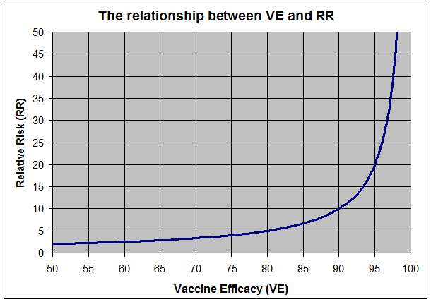
Or, for those of you who are more visual, here’s a graph of which RR values correspond with which vaccine efficacy values.

How Relative Risk values convert to Vaccine Efficacy values. Keep in mind that Relative Risk can vary from 1.0 (no difference in disease rate between groups) to infinity (disease in the unimmunised but complete protection of the immunised), so conversion to the 0-100 Vaccine Efficacy scale requires a steep curve as VE approaches 100%

So imagine a vaccine with a Vaccine Efficacy value of 50% – which corresponds to RR=2, or the unvaccinated having twice the rate of disease of the vaccinated. If you study two equally sized groups of people that did and did not receive said vaccine, in an outbreak you’d expect the unvaccinated to have twice as many cases of the disease. But if the unvaccinated group was twice as big as the vaccinated, you would expect to see equal numbers of cases in both groups, and similarly you would expect to see more cases in the vaccinated if they outnumbered the unvaccinated by more than two to one. So although the vaccinated are at a reduced risk of catching the disease, if they outnumber the unvaccinated by more than the risk ratio for that vaccine, we should actually expect to see more cases of the disease, by number, in the vaccinated, despite a lower risk per person. If you’d like to play around with how the maths works, below is a simple calculator into which you can put a vaccine efficacy value and a vaccination rate for a hypothetical disease, and it will tell you how many cases you’d expect to see in the vaccinated for each case in the unvaccinated, assuming equal exposure to disease between the immunised and unimmunised groups. Have a play around and see if you can change the parameters to get more cases in the vaccinated group than the unvaccinated group. If you’d try it with some real VE values, on the high end of the scale one dose of MMR vaccine has a VE of at least 95% against measles, and on the low end of the scale the CDC’s mid-season estimate of the influenza vaccine for the 2013-2014 ‘flu season was 61%.

For an outbreak of a vaccine preventable disease with

A vaccine efficacy of: %

A vaccination rate of: %

For every cases of disease in the unvaccinated, we should expect to see 60 cases of the disease in the vaccinated.

In summary, vaccines reduce the risk of contracting diseases, they do not eliminate this risk entirely. Even though the unimmunised may be at a considerably higher risk of contracting a disease, because they make up such a small sliver of the population, we should not be surprised to see them representing the minority of cases during outbreaks of vaccine-preventable diseases. As explained above, this is not a challenge to the effectiveness of vaccines, but instead is to be expected in populations where the vaccinated outnumber the unvaccinated.

While not discussed here, ‘vaccine effectiveness’ refers to results from actual outbreaks in general populations, while ‘vaccine efficacy’ is used when assessing a vaccine in a highly controlled manner, which can have reduced generalisability to broader populations
P.s: Someone has asked to use this post and the tools herein elsewhere. If you would like the HTML code for the calculators (they’re pretty simple) just leave a message or contact me @kill3rTcell and I’ll flick them to you.
These equations and others for assessing and quantifying real-world effectiveness of vaccines are described in this paper from the Bulletin of the World Health Organisation.
Posted in Uncategorized, Vaccination | Leave a comment

Protected:

This content is password protected. To view it please enter your password below:

Posted in Vaccination | Enter your password to view comments.

An analysis of Rose Lalonde’s MEOW DNA code

My apologies to my regular readers, this is somewhat of a divergence from my usual subject matter – just click ‘back’ to get to previous posts on the top left above this article if you want to read one of my usual articles about vaccines, immunity or whooping cough. Unless you are familiar with Homestuck (a short, happy story about some friends who play a videogame together while being pestered by internet trolls) this post is unlikely to make any sense to you, and unless you are beyond Act 4 of Homestuck, contains spoilers.

The background: On this page of Homestuck we see a page of Rose’s MEOW journal, with a 952 character long excerpt in the Journalog below it. JournalogEach of these letters represent one of the four DNA nucleotide bases, apparently M=G, E=C, O=A and W=T  (which I’ll refer to as ‘MEOW = GCAT’ for short), as we discover in this part of the flash [S] Jack: Ascend:

MEOWGCAT I have not been able to find online an analysis that goes suitably in-depth into this code to satisfy me, so I went ahead and performed one myself. I looked as hard as I could for any biological relevance of the DNA sequence, as well as for a hidden message in it, not only with the key MEOW=GCAT, which [S] Jack: Ascend indicates is the correct substitution, but also with all twenty four possible nucleotide base substitutions – and all of those can be accessed/downloaded below for anyone who wishes to look for messages in there themselves. But before you do, I don’t believe there is any hidden information  in Rose’s MEOW code to be found.

I investigated a few theories of how Hussie might have come up with the string of letters that makes up the MEOW code, and I found evidence for what was possibly the least satisfying one of all… that Hussie just made up a random sequence of the letters M, E, O and W using his trademark keyboard-hitting:

AH: Hit the M, E, O and W keys nearly 1000 times to make up Rose’s Journalog

I’ve tried to generate a random, long string of DNA bases myself in the past, and quickly resorted to copying and pasting chunks of text I already typed out in order to finish the job. Hoping that Hussie might have got bored mid sequence and done the same as I had, I put the MEOW code into a repeat finder, and found it was indeed full of repeated sequences.

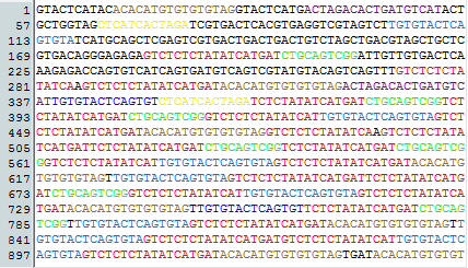
I took a day to identify all of the repeated sequences and colour-code them. The image below is the MEOW code, where every time the same sequence appears, it is the same colour. As you can see up until about 270 letters into it there are no repeats (each coloured sequence appears only once until then). From that point on though, the sequence is basically just a wall of colour, indicating it’s made almost exclusively of strings of letters we’ve seen before:

The MEOW code, presented as MEOW=GCAT. Each time the same sequence appears, it is labelled in the same colour. Generated using Serial Cloner v2.6.1

The MEOW code, presented as MEOW=GCAT. Each time the same sequence appears, it is labelled in the same colour. Numbers on the left correspond to the adjacent letter’s place in the sequence. Annotated using Serial Cloner v2.6.1

Over 70% of the MEOW code is strings of letters which have previously occurred in it, and strings which are so long that the likelihood of them having occurred twice by chance in a sequence of this length is less than 1/10,000.

I’m sorry to say it, but the MEOW code really does just look like it was created by whacking those four keys about three hundred times, then copying and pasting bits of what was already typed out to finish it off.

What sold it for me was when I realised the very final repeat of the brown sequence follows the letters ‘TGAT’, whereas previously it always followed the red sequence. I then realised that TGAT are the last four letters of the red sequence – indicating that someone had incompletely copied and pasted part of both sequences to make this very final addition to the end of the code:

That thing I just said

That thing I just said

So the code is exactly what you’d expect to see if someone tried to enter a random sequence, but got bored part way through and just copied and pasted chunks of what they’d already typed in to finish it off.

I’ve got to give it to Hussie though, despite over 70% of the code being repeated sequences, he actually did a pretty good job of randomising their order to make it less obvious, as you saw from the distribution of colours throughout the previous image.

What follows are my attempts to find any information in the meow code. I don’t believe there’s anything to find, but it was worth a try to see if there was anything interesting there.

So how might information be encoded in the sequence? Well the two most promising ways would be:

  1. The journal excerpt could be a chunk of a DNA sequence from a real creature. The code is used in canon to create Bec, so it would be pretty cool if it were actually part of a dog genome sequence, right?
  2. Or, a message may be written in there. Yes, written. Not in M, E O and W, or the DNA bases G, C, A and T, but in the protein encoded by the DNA sequence. Groups of three DNA bases encode individual amino acids. There are twenty amino acids in the DNA code and each is represented by a letter of the alphabet (the six letters not used in this code are B, J, O, U, X and Z). In this way it is possible to encode whole sentences in a DNA sequence (in reality those strings of amino acids are proteins, but on a computer it’s a cool way to encode some information).

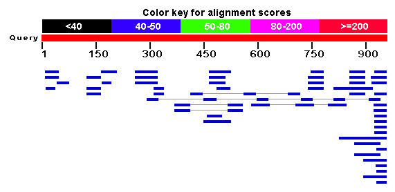
So, might Hussie have included part of an actual gene in this sequence? Thankfully, the National Center for Biotechnology Information allows us to submit a query DNA sequence, and it will search through the biggest assembled database of DNA sequences (including dog genome sequences) for ones that match the query. So what do we get when we perform a search for sequences matching the MEOW = GCAT sequence using the BLAST algorithm?

The DNA sequence of MEOW=GCAT was entered into a BLASTn algorithm search. The sequence is represented above in red, and each of the matching sequences below are in blue, indicating that they have a very poor alignment score. The grey lines indicate where ‘matching’ sequences needed to be broken apart in order to align the query sequence.

Above is the graphical output of the BLASTn algorithm. Each of the matching sequences are shown in blue, indicating their poor alignment score (a score of only “40-50” according to the key; perfect matches are typically red). Each rectangle is a ‘matching’ sequence. Not only do they score poorly, but you can see each rectangle only covers a small portion of the sequence (where they are under the sequence indicates which part they align with).

Disappointingly, the DNA sequence encoded in Rose’s journal does not match any sequences from the largest accessible database. All we see are small chunks of the sequence that happen to match (with a poor score) short chunks of other sequences – exactly what you’d expect if you entered any long, meaningless, made-up DNA code.

However, on the off chance that MEOW=GCAT was not the right key, I performed BLASTn searches for all possible MEOW to G, C, A or T substitution variants. None of the reports finds significant similarity with any sequences in the database, but if you’re curious you can view each of the BLASTn reports below (note, the ‘ACGT’ link means the code I used was MEOW=ACGT, while in ‘GCAT’ I used MEOW=GCAT, etc.)

ACGT ACTG AGCT AGTC ATCG ATGC CAGT CATG CGAT CGTA CTAG CTGA GACT GATC GCAT GCTA GTAC GTCA TACG TAGC TCAG TCGA TGAC TGCA

Okay,so none of the possible MEOW/GCAT substitutions matches any archived genome, gene or RNA sequences. But what about the second information-hiding possibility I mentioned? Could a message be written in there? Well, three DNA bases encode a single amino acid, which means there are three reading frames depending on which base you start at, and three more if you choose to decode the sequence backwards. So in short, any DNA sequence could potentially encode six different strings of amino acids. Here are the six encoded by Rose’s MEOW code, where the substitution MEOW=GCAT is used. Each row is one of the six possible reading frames:

Tcl Printer Document

The DNA sequence in Rose’s Journal, where M=G, E=C, O=A and W=T. Below the sequence are the amino acids encoded in it. The top three rows of amino acids are reading frames 1, 2 and 3. The bottom three rows (reading frames -1, -2 and -3) should be read in reverse. Generated using ‘A plasmid Editor v2.0.47′

Disappointingly, I can’t make out any significant words, let alone sentences, in this code.

However, on the off chance that Hussie may have encoded something, but with a key other than MEOW = GCAT, I did the same with all 24 possible MEOW to G/C/A/T substitutions. I could not make out any messages in them, but if you want to see for yourself, each can be viewed in PDF format in these links. The name of each file indicates what key was used (eg. ‘GCAT’ uses the key MEOW = GCAT).

ACGT ACTG AGCT AGTC ATCG ATGC CAGT CATG CGAT CGTA CTAG CTGA GACT GATC GCAT GCTA GTAC GTCA TACG TAGC TCAG TCGA TGCA TGAC

In summary, I have not been able to find any real genetic information, or coded messages in Rose’s MEOW Journalog excerpt, and I believe this is because there are none there – Hussie likely just created the code randomly and finished it by copying and pasting bits of it he already typed out, as evidenced above.

Fellow Homestuck fan, I’m sorry the outcome of this analysis wasn’t more exiting. I was earnestly expecting some message from Hussie in the amino acids, or failing that, discovering it was part of a dog gene, and was a little disappointed on finding the truth, and hope this post wasn’t that much of a let-down. But hey, you wouldn’t be a Homestuck fan if you weren’t able to read walls of text with the occasional picture, only to be disappointed by it, right?

If you are interested in performing any of these analyses for yourself, or taking them further, please find the MEOW code here. Each of the tools used in the analysis are free and linked where mentioned in the post. The colour codes in the ‘repeats’ analysis have been simplified, there are four copy/paste variants of the red sequence, and two each of the blue and brown sequences, where they seem to have been copied incompletely prior to pasting. A version with multiple shades of these colours to illustrate this can be viewed here. Feel free to ask questions in the comments :o)
Posted in Homestuck | Tagged , , , , | Leave a comment

Waning pertussis immunity in England/Wales, and bad reporting in Australia

A new study has come out from the UK, assessing the case for the National Health Service adding an adolescent booster of the pertussis vaccine to the schedule.

I’m sure many of us in countries such as Australia, New Zealand, US and some European countries may be surprised to hear that this routine (to us) shot is not on the NHS schedule, but they did pioneer an advanced infant schedule which saw increased protection from pertussis in this age group. As you can see from the figure below, introduction of 2, 3 and 4-month pertussis shots onto the UK schedule in 1990 was followed by an absolute decrease in infant pertussis cases in the < 3 mo, 3-5 mo and 6-11 mo age groups (top graph), and following this change in the schedule, the < 3 month age group went from representing under 15% of cases in infants to 40-50% of these cases.

Age-group distribution of case-patients <12 months of age in pertussis case-notification reports, (Top) by total case patients and (Bottom) by proportion of age, England and Wales, 1983-2009. Red, < 3 months of age; Blue; 3-5 months of age; Black, 6-11 months of age. Source.

However, as well as this clear evidence of effectiveness in the young, this earlier study also found pertussis to still be maintained in the adolescent and adult populations, where pertussis vaccine coverage is poor. As I’ve discussed before, duration of protection from the currently used acellular pertussis (aP) vaccine is estimated to last from four to twelve years, with recent result indicating the evolution of the bacterium may be contributing to early waning of immunity. While some countries have controlled pertussis in the adolescent cohort with later booster doses, the UK health authorities are currently considering adding such a booster to their schedule.

The authors of this new study set out to assess what proportion of persistent coughing illness in adolescents in the Thames Valley area was being caused by pertussis infection, and how prevalent previous encounter with pertussis may be in this cohort. The study authors considered adolescents with persistent cough that had evidence of previous pertussis immunisation, and the correlation between pertussis incidence and time since last booster was much like what we’ve seen previously:

Laboratory confirmed pertussis in children presenting with persistent cough in primary care after receiving preschool pertussis booster vaccination (n=224). Error bars represent 95% confidence intervals

Laboratory confirmed pertussis in children presenting with persistent cough in primary care after receiving preschool pertussis booster vaccination (n=224). Error bars represent 95% confidence intervals

The increased risk of pertussis infection in children who received pertussis immunisation over seven years ago is consistent with previous findings that immunity wanes over this time period, to the extent that non-recent vaccinees are at similar risk of catching the disease to unvaccinated controls.

Keep in mind that this study only included immunised children, so there is no quantification of absolute risk relative to the unimmunised, though, as discussed above, that absolute protection has been demonstrated. That said, these results do answer the researchers’ question of how risk of pertussis increases with time in this age cohort not covered by a booster shot.

The authors also assessed the number of times six patients coughed in a 24 hour period. Two coughed around 200 times, three around 500 times and one 1600 times, and on the basis of this the authors reinforce that, despite the established protection of recent vaccine-recipients from more severe pertussis disease, the once aP vaccinated can still suffer relatively severe symptoms on eventual infection. This is not the first time the authors have reinforced in a paper the importance of pediatricians considering pertussis as a potential diagnosis in cases of persistent cough, and hopefully it gets this point across to pediatricians who keep track of the literature.

However, the big, headline-grabbing result from this study is that, of cases of persistent cough in previously pertussis immunised children presenting to primary care physicians, 20% had evidence of recent pertussis infection. While this prevalence of pertussis is indeed a matter of concern, it is worth pointing out several nuances that do not seem to have made it into the media as well as this soundbite:

  • The rate of pertussis in this age cohort found in this study is decreased compared to the rate seen prior to introduction of the preschool pertussis booster
  • The cohort with the greatest infection rate (by 2-3 fold) in this study was one in which aP-induced immunity is known to have largely waned

While it’s quite clear that the anti-immunisation lobby is going to jump all over selective quotations of this paper as evidence the pertussis vaccine is useless, in actuality these results indicate less pertussis infections in this age group since the introduction of the preschool booster (about half of the previous rate, despite over a third of this study occurring during a pertussis epidemic), and that the preschool booster has pushed back the time of immunity waning by several more years.

Oh, and another outcome of these results that could lead to unwarranted excitation of the anti-immunisation lobby is misreporting. From Medical Observer on this study yesterday:

Screenshot from the Medical Observer article in question

Furthermore, the risk of pertussis was more than three times higher in children who had received the pertussis booster less than seven years previously.

Which you might notice contradicts the results of the study, and the BMJ‘s plain language Research News‘ article on the study which I suspect is were many news outlets got their interpretation of the study from:

The risk of pertussis was more than three times higher (21/53; 40% (26% to 54%)) in children who had received the preschool pertussis booster vaccination at least seven years before presenting with cough compared with those given the booster less than seven years previously (20/171; 12% (7% to 17%)).

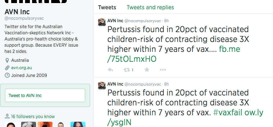
With such a severe mis-reporting of the study’s results, it’s only a matter of time until the anti-immunisation lobby, which regularly repeat the lie that vaccines increase your susceptibility to the disease and who never bother to follow up on news reports to see whether their claims are backed by the primary sources, touts this article as evidence of its claims.

Oh wait, it’s already happening.

Yup

Screenshot from the official twitter page of the Anti-Vaccine lobby group, the Australian Vaccination Sceptics Network.

Screenshot from the Australian Vaccination Sceptics Network twitter page. As you can see the, this anti-vax group have quoted verbatim Medical Observer‘s headline: “Pertussis found in 20pct of vaccinated children“. Great choice of wording there, MO.

Update: As of “3.34pm 26 June 2014“, Medical Observer have ammended their article to correct the highlighted error, but have maintained the misleading click-bait headline “Pertussis found in one fifth of vaccinated children“.

Posted on by kill3rtcell | 1 Comment

The Anti-Vaccination Network’s talk at the Healthy Lifestyle Expo

Yes, She Actually Said That

On the 25th of May 2014, the spokesperson for the Australian Vaccination Skeptics Network (AVSN, formerly the Australian Vaccination Network), Meryl Dorey presented a talk, Vaccinations – Do They Really Save Lives? at the Healthy Lifestyle (You Can Heal Yourself) Expo in Caloundra, Queensland.

If you’re familiar with the AVSN, you know they’re an anti-vaccine group that do not provide reliable information on vaccines, and in fact disseminate misleading, misrepresented and incorrect information about vaccination which engenders fear and alarm, according to the public statement the New South Wales Health Care Complaints Commission put out about them.

You may have heard of their spokesperson and public officer, Meryl Dorey, who has no qualifications in immunology, microbiology, medicine or vaccinology and who for over a decade has been the major voice behind most of the incorrect information  put out by the group. This talk was no exception.

In reading the transcript there were three particularly egregious sections that showed a  complete ignorance of immunology, and I must admit to enjoying falling down the rabbit hole of reading the broader history behind all three of them. So without further ado, Meryl Dorey’s own words, followed, of course, by referenced descriptions of the evidence around the claims.

Questioner: “Hi Meryl, thanks for coming along, Lee is my name. Uh I once worked with a bloke who had a severe allergy to peanuts, sesame oil and seeds. He had one mouthful one day, grabbed his throat and dived to his pocket for a pill. It was that quick, it fixed him but I only recently read something about the alleged cause of it but I don’t fully understand it, can you explain it in simple terms, something about the oil being used as a carrier to keep it effective for longer. Sorry on the um, um placebo, uh can you explain if I’m allowed to explain why, how many studies on vaccination effectiveness use uh adjuvants and you know, thiomersal as the placebo.”

Meryl Dorey: “Um, the peanuts, peanut oil we believe has been included in vaccines since the late 1960s. I am 56 years old, I was born in 1958. I never knew anyone with peanut allergies until I started having my own children. So for a long time, 30 years, I never knew about peanut allergies, now you don’t know anyone who doesn’t have them. And um, there’s an excellent book that I can recommend called “The Peanut Allergy Epidemic”, Heather Fraser I think is the name of the author, and she discusses it. Um they have evidence that peanut oil is used as a carrier in many vaccines and to this day it’s used in many vaccines. And because, probably too long to go into, but because vaccines are intended to alert your immune system to the fact that there is an invader in there, and if you attach a peanut oil, let’s say, or an oil, a lipid of some kind to uh, a virus or a bacteria and then you inject it into the body, the body can look at that and say, okay I see measles and I know that measles is an enemy – this is the theory and I don’t know if it’s true but this is the theory – I know that measles is an enemy and attached to that measles I’ve got this weird oil. Now I’ve got something in my body that looks just like that, uh, I have all of this myelin wrapping around my nerves, and other oils that my brain uses, so I’m going to attack this because it’s attached to measles and when I see this oil in something else, in something that I eat or in my body I’m going to attack that too. So I’m going to develop a peanut allergy, I’m going to develop anaphylaxis and I can die after peanuts.

The interesting thing is that in Asian countries, some Asian countries, peanuts are not common but um sesame seeds are, they use sesame seeds in their food, so in those countries we’ve been told that they use sesame oil in their vaccines and just the way that we have these peanut oil allergies, they have sesame allergies as well. So whether there’s proof of that, I don’t know, but it’s certainly interesting and it should be studied.”

The claim that peanut oil has been included in vaccines since the sixties is downright false, with peanut oil not in any vaccinations on the Australian or American schedules.

Only on anti-vaccines sites is this claim made. In lieu of any proof that vaccines currently contain peanut oil, the authors of those pages point to studies performed with experimental vaccines containing peanut oil based adjuvants, most citing papers detailing the development and eventual abandonment of ‘adjuvant 65-4’. As with any water-in-oil emulsion-based adjuvant, vaccine antigen was made up in water and an emulsifier used to suspend it in oil. However, unlike mineral oils which the body has difficulty clearing (and as such are not licensed for use in human adjuvants), the use of peanut oil in adjuvant 65-4 meant the oil was completely metabolisable. Contemporary studies showed a positive safety profile and duration of immunity in humans and animals for an experimental adjuvant 65-4-containing influenza vaccine, leading to a United Kingdom licence in 1973. However, the US requirement that the oil mannide monooleate be used instead led to reduced efficacy and saw the eventual abandonment of the entire adjuvant 65-4 vaccine project.

Similarly, sesame oil has been used experimentally in emulsionbased adjuvants.

The entirety of the evidence for the claim of peanut and sesame oils being in modern vaccines, as found on anti-vaccine sites, consists of links to early studies and reviews of peanut- and sesame-oil adjuvants, cited as somehow being proof they are included in current vaccines. In reality, the only emulsion-based adjuvants licensed for use in humans use the oil squalene.

A common trope to try and worm around this lack of evidence, used by many American-based anti-vax sites, is to say that because peanut oil is classified ‘generally regarded as safe’ (GRAS) by the United States Food and Drug Administration it does not need to be mentioned in the labelling of vaccines. However, a complete list of substances involved in vaccine production reveals that no peanut products are used at any stage of the process.

So Ms Dorey’s claim is presented not only without evidence but on investigation is found to be baseless, though oft-repeated in online anti-vaccine conspiracy websites.

But what about this part?

“And because, probably too long to go into, but because vaccines are intended to alert your immune system to the fact that there is an invader in there, and if you attach a peanut oil, let’s say, or an oil, a lipid of some kind to uh, a virus or a bacteria and then you inject it into the body, the body can look at that and say, okay I see measles and I know that measles is an enemy – this is the theory and I don’t know if it’s true but this is the theory – I know that measles is an enemy and attached to that measles I’ve got this weird oil. Now I’ve got something in my body that looks just like that, uh, I have all of this myelin wrapping around my nerves, and other oils that my brain uses, so I’m going to attack this because it’s attached to measles and when I see this oil in something else, in something that I eat or in my body I’m going to attack that too. So I’m going to develop a peanut allergy, I’m going to develop anaphylaxis and I can die after peanuts.”

Ms Dorey goes on to tell the questioner that the immune response generated against peanut oil in vaccines cross-reacts with myelin in the brain. While molecular mimicry – the phenomenon of an immune response against one molecule leading to an immune response against another similar molecule – is considered as a viable hypothesis for the development of demyelinating diseases, a literature search for the terms ‘peanut myelin’ fails to turn up any research demonstrating co-occurrence of peanut allergies with demyeinating disease, or any molecular evidence of immunological cross-reaction between antigens from either of these sources. Not only are peanut products not used in vaccine production, but there does not appear to be any evidence linking peanut allergies to demyelinating diseases as Ms Dorey claims.


The next excerpt contains a claim Ms Dorey has been making for a while now.

“And that sounds all well and good except we have known for over 70 years that the presence of antibodies do not mean that you are immune to the disease, in fact all it means is that you’ve been exposed because many people can have absolutely no antibodies to a disease and be immune to it and many other people can have very high levels of protective antibodies and not be protected. Um this is an obituary for Dr Merrill Chase, a man Merrill, and he is called the father of modern immunology and he discovered in the 1940s that it is not antibodies alone that protect us. The immune system is incredibly complex and uh a balanced, a wonderfully balanced system that, that, has been developed over millions of years in order to help protect us from the environment; from toxins, from diseases in the environment. And what Dr Chase found is that antibodies are only a small part of that process, they’re not the be all or the end all and that can explain why people who have very high levels of antibodies still get the disease. So the whole purpose of vaccination is to create these antibodies, to cause our bodies to create these antibodies, and yet that doesn’t mean that we’re going to be protected at all.”

This is a claim Ms Dorey has made before,  specifically that vaccines are designed primarily to get B cells to produce antibodies against vaccine antigen, but because there exists another arm of the adaptive immune system (T cells, the cellular mediators behind Merrill Chase’s observations), and because antibodies are not an absolute marker of protection, vaccines therefore induce ineffectual protection.

First off, it must be pointed out, T cell responses induced by vaccines on the schedule have been  researched and characterised.

However, what is particularly humourous about this claim is that knowledge of T and B cell interactions has actually been incorporated into vaccine design since even before the AVSN was founded. The conjugate Haemophilus influenzae type B vaccine, licensed in the United States since 1987, includes a protein carrier specifically chosen for its ability to stimulate T cells, to take advantage of the improved immune response that occurs when cell-mediated immunity and B cells cooperate.

As for Ms Dorey’s claim that antibodies are no indication of whether you’re going to be protected at all, specific antibodies induced by vaccination are a quite effective indicator of the level of protection afforded to vaccine recipients. Decades of work show that by observing levels of antibody induced by vaccination and correlating it to disease outcome, it is possible to define a threshold of induced antibody above which an individual is at drastically reduced odds of developing disease on subsequent exposure. However, reduced risk of disease with high levels of antibody is not the same as no risk; the fact of the matter is that high levels of specific antibody correlate with a lower chance of catching disease, and better clinical outcome in those ‘breakthrough’ infections. However, the fact that small proportions of people may come down with disease despite being above an experimentally determined threshold does make Ms Dorey’s comment, “So the whole purpose of vaccination is to create these antibodies, to cause our bodies to create these antibodies, and yet that doesn’t mean that we’re going to be protected at all” technically true, if used entirely deceptively.

It’s worth pointing out that Ms Dorey’s dismissal of this work is not limited to rejecting the existence  of an antibody-prognosis correlation simply because it is not absolute (the Nirvana logical fallacy), in fact she denies any immunological role for antibodies, outright describing them as “Peripheral to protection”, showing ignorance of decades of research that demonstrates protection mediated by direct transfer of antibodies, transfer of antibodies through breastfeeding and more recent highly specific mAb therapies.

Although one may suspect innocent oblivion on reading Ms Dorey’s diatribes that specific antibodies are nothing more than a marker of previous exposure, in the same post she cites as sources:

A review which describes in detail the protective levels of antibodies induced by vaccines, the evidence of antibodies as direct mediators of immunological protection and which, when it comes to the discussion of T cell responses, describes as “obvious” that “antibodies in sufficient quantity are the predominant protective correlate“.

A textbook chapter, which describes the importance of antibodies in mediating protection and paints a picture of T cell aspects of the response as primarily supporting antibody production.

A research paper in which mice whose B cells were unable to secrete antibody succumbed to an infection that wild-type mice survived (the authors go on to describe this antibody mediated protection as “critical” to surviving the infection) – an observation entirely incompatible with the antibodies being “peripheral to protection”.

Ms Dorey cites each of these as a source and yet continues to spread counter-factual information that they each refute, making it impossible to attribute this campaign of misinformation to mere ignorance. It seems Ms Dorey is either sufficiently scientifically illiterate to not realise the papers she cites for support are incompatible with her contentions, or she is deliberately deceiving the public about the efficacy of vaccines and basic cellular and molecular functions of the immune system.

In summary, Ms Dorey states in the quote:

1 – That mechanistically, vaccines cannot work – apparently unaware that the mechanism she states they omit has been harnessed for decades to improve vaccine efficacy; and

2 – That a major cellular mechanism by which protection through immunological memory is maintained is pure fiction, despite over a century of research demonstrating its reality.

Such egregious misinforming of the public simply does not constitute appropriate behaviour for a health care provider.


Perhaps the best part of the talk though:

“Questioner: Hello, my name is Melanie. Thank you for your talk, Meryl, it was really informative. I did some research recently about human foetus cells being used in vaccines and I got some information off you. My question is – and as you know a lot of aborted foetuses are sold to the pharmaceutical industry each year – if the mother is vaccinated of that foetus will any of that carry into the foetus negative or positive? [inaudible]

Meryl Dorey: I just want to make sure I have that question right. The question is uh, the foetuses of mothers, mothers who abort their foetus, hospitals sell those foetuses to vaccine and drug companies, right? Okay, and that’s true. Would vaccines that the mother have received actually be uh the antibodies from those vaccines be in the baby and I’m saying yes, they would because um, the aborted foetuses that were used to make the rubella vaccine and the other vaccines that use aborted foetal tissue, chicken pox and others. The reason they were selected is because their mother had had rubella or chickenpox and the antibodies were present in the foetus. So yeah, they would be I think, as far as I know.”

Meryl Dorey tells this questioner that a black-market trade of foetuses aborted from mothers infected with vaccine-preventable diseases exists, to supply the raw materials for making more vaccines against those diseases.

This is entirely false.

“…mothers who abort their foetus, hospitals sell those foetuses to vaccine and drug companies, right? Okay, and that’s true.”

Australian law prohibits trade in human body parts, with the National Health and Medical Research Council’s Ethics and the exchange and commercialisation of products derived from human tissue deferring to section 4.1.13 of the National Statement on Ethical Conduct in Human Research (2007) which states “There should be no trade in human fetal tissue.

Further, foetuses are just not required for the ongoing production of vaccines.

In the 1960s two cell strains (WI-38 and MRC-5) were derived from legally medically-aborted foetuses. These cell strains simply need to be given the right conditions to survive and replicate; the progeny of those cells isolated in the ’60s are still used and dividing today in laboratories around the world. Because viruses need to infect living cells to replicate, the viral components of Hepatitis A, Rubella, Varicella, Adenovirus and Rabies virus vaccines are grown in those two cell strains. Vaccine production just does not need a periodic top-up of fresh fetuses as Ms Dorey says is provided by her fictional fetus-trade.

“The reason they were selected is because their mother had had rubella or chickenpox and the antibodies were present in the foetus.”

The cell strains were not chosen to grow these viruses because the mother was infected with them (firstly, there are two strains and five viral species cultured in them), they were chosen simply because they supported growth of those viruses. The mothers’ infection history and whatever antibodies they were passing on to their fetuses are entirely irrelevant to this. The presence of antibodies against the virus in question should be expected to be a hindrance to their culture, and could be expected to interfere with the action of the vaccine should they make it into the final product.

Ms Dorey’s talk shows ignorance of some basic concepts in immunology (such as what antibodies are, and a role of T-B cell interactions which has been incorporated into vaccine design longer than the AVSN has existed) and vaccine production (featuring a description of virus culture so fantastical it breaks Australian law). What is worse, Ms Dorey’s previous use of references that so neatly contradict so many of these claims makes it very difficult to believe these are simple naive mistakes.

No group that so radically and deliberately misinforms the public deserves the official status of healthcare provider that the Australian Vaccination Skeptics Network has.

Posted in Australian Vaccination Network, Vaccination | Tagged | 10 Comments

Why as a researcher in medical science I oppose Hockey’s billions for medical research

Posted in Uncategorized | Leave a comment

Whooping cough bacterium loses expression of a key vaccine target

This post is a discussion of the April 2014 study showing Australian pertussis bacteria to be losing expression of the vaccine target pertactin. The post describing the evidence that the pertussis vaccine does not increase susceptibility to parapertussis is here.

Finally, more information has come out about the proportion of Whooping Cough bacteria that are losing expression of a protein targeted by the current vaccine.

The background it relatively simple: from the 1950s a vaccine against whooping cough was used that contained whole, killed cells of the bacterium responsible for the disease, Bordetella pertussis. Although the biggest studies to date have failed to support the claims, contemporary fearmongering led to an increase in rejection of the vaccine and in lawsuits filed against its manufacturers and a subsequent shortage of the shot. Part of the solution to the drop in public confidence was the development of a vaccine that only contained several key proteins of the pertussis bacterium, the acellular pertussis (‘aP’) vaccines. In Australia, the whole cell vaccine was fully phased out of routine childhood immunisations in the early 2000s, with an aP shot containing three pertussis proteins currently on the childhood schedule.

Last year, an American report noted that of 12 clinical isolates (that is, samples of the bacterium obtained from sick patients) of Bordetella pertussis they tested, 11 had lost expression of Pertactin (Prn), one of the proteins included in modern formulations of the aP shot. Given that the version of the shot subsidised for the childhood schedule only includes three pertussis proteins, one of which is Prn, there is the possibility that this loss, if widespread, may have serious implications for the effectiveness of the immunity induced by the vaccine. As I predicted in my coverage of that paper, Australian researchers have since addressed the questions of how frequent these Prn-negative strains are and how recently they’ve arisen. In this post I will try to summarise for non-biologists the April 2014 paper, Rapid Increase in Pertactin-deficient Bordetella pertussis Isolates, Australia.

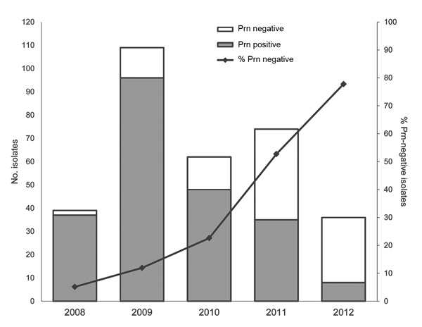
The juicy bits of the paper are summarised in their Figure 2 (below) which shows how many isolates from each year did or did not have detectable Prn expression (the bars) and what percentage overall were negative (the line).

Slide 1 The authors report only being able to detect Prn-negative strains from 2008 onward, with a sharp rise that culminates in ~80% of isolates lacking detectable Prn protein in 2012. This suggests that there may be a selective advantage for Prn-negative strains of B. pertussis.

In order to determine whether this was a single chance event which gave one strain superiority, or possibly a more widespread loss of Prn protein expression in the wild B. pertussis population, the researchers decided to sequence the gene that normally encodes for Prn protein to see why it had been lost.

Interestingly, the team discovered that there were multiple reasons for the block in protein production. 80% of Prn-negative isolates had insertion sequences – pieces of mobile DNA, sort of like mini-viruses that jump through the genome – smack bang in the middle of the gene, stopping production of the protein. Two of the 96 Prn-negative isolates seemed to have complete deletion of the gene. Sixteen of them had completely normal Prn genes, indicating some other factor regulating production of the protein must have been altered. All of these mechanisms again differed from those reported by researchers outside of Australia who reported Prn-negative pertussis isolates. In short, there have been multiple roads to Prn-protein negativity taken by the pertussis bacteria, which suggests there has been a strong selective pressure on it for so many ways of losing the protein to have spontaneously arisen in the B. pertussis population.

So what does pertactin loss mean? From the vaccine effectiveness standpoint, if we assume near-complete loss of Prn expression, then our most-used vaccine now effectively only targets two pertussis proteins, PtxA (the active subunit of the pertussis toxin) and Fha (filamentous haemagglutinin – used by the bacterium to stick to things, such as our respiratory tract). As I described last time, the biggest prospective, randomised, placebo-controlled clinical trials of aP vaccines that only target these two proteins showed an effectiveness of 59-69% which equates to a 2.4-3.2 greater rate of pertussis disease in the unvaccinated controls compared to those who got the shot. While these figures are indeed suboptimal when compared to many other vaccines on the childhood schedule, it shows that targeting PtxA and Fha alone can mediate protection from pertussis.

What about the effect of Prn protein loss on the bacterium? Well, the authors point out previous work that found Prn-mutants were less able to colonise mouse respiratory tracts, but those that managed to colonise were more able to invade the cells lining the tract and persisted for longer. The authors also point out work that found Prn-deficient mutants grew faster in the lab than the Prn-expressing bacteria they were derived from, but it’s unclear whether that has any relevance to an infection scenario – the fewer other proteins the bug has to produce the more resources it can dedicate to reproducing, but the loss of a protein that helps colonisation may not be worth it in an infection.

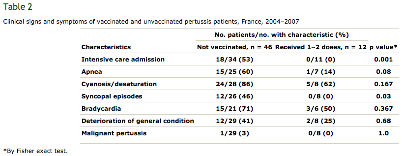
Lastly, covering the relevance of Prn loss to clinical outcomes, the authors discuss a recent French retrospective study which compared the health outcomes of infants that had infections with Prn-positive or -negative B. pertussis. Prn-negative infections were found to induce no worse clinical symptoms than Prn-expressing ones, consistent with earlier mouse work from the same group which found Prn-negative isolates to be no more lethal. The French study also found infants with Prn-negative infections spent on average four days less time in hospital than Prn-positive infections, suggesting the loss may negatively impact the bacterium’s ability to cause severe disease, though the sample size was not big enough for the difference in hospital stay times to reach statistical significance. Lastly, the French group found that, regardless of Prn expression, immunisation against pertussis was significantly associated with protection from the worst of the disease – just have a look at their Table 2, below:

In summary, Australian Bordetella pertussis bacteria are increasingly losing expression of pertactin protein. This apparently global trend by the species, with multiple mechanisms of loss (and even by the closely related species Bordetella parapertussis), is consistent with this being a response to vaccine-induced selective pressure. However, both human and animal data indicate that Prn loss does not worsen disease, and shows that the pertussis vaccine is still the best broadly available protection from whooping cough.

The long-term solution to the problem of the bacterium slowly evolving around the immunity induced by the vaccine is a new, more broadly-acting vaccine. And, as I’ve discussed before, there are several whole cell variants in various stages of testing being reported in the literature. In the short-term, people should get their adult boosters and protect themselves from pertussis disease. I got mine last year, making sure to ask for the five-component variant (Adacel in Australia), in the hope of that extra protection.

It’s quite clear now that there’s a problem, with the whooping cough bacterium gradually accruing mutations which should afford it some protection from immunity induced by the vaccine. I hope in the coming months we will see some sensible action by the Australian Technical Advisory Group on Immunisation, as I don’t want to see what the national pertussis statistics may look like in a decade if this problem is not addressed soon.

Posted in acellular pertussis, Acellular pertussis vaccine, Bordetella pertussis, Pertactin, Pertussis, Pertussis immunisation, Pertussis vaccination, Vaccination, waning immunity, waning pertussis immunity, Whooping cough, whooping cough immunisation, whooping cough vaccine | 4 Comments

Death threats and threats of harm incited by the Australian Vaccination Network (NSFW)

Posted in Uncategorized | Leave a comment

‘Vaccine resistant’ pertussis – in a way, yes. In a more accurate way, no.

This post is a discussion of the February 2013 report from Philadelphia that the pertussis bacterium may be losing expression of the vaccine target protein pertactin, and the implications of such a loss. A discussion of the April 2014 paper that found most Australian pertussis bacteria have lost pertactin expression can be found here.

A recent letter to the New England Journal of Medicine is making the news rounds online. With the title Pertactin-negative variants of Bordetella pertussis in the United States (full text here) it describes the findings that some of the whooping cough bacteria (Bordetella pertussis) in the US appear to have stopped expressing one of the proteins targeted by the vaccine. With headlines like “Whooping cough may be becoming resistant to vaccines” and “Some Whooping Cough Strains Now Outsmarting Vaccine” circulating in response to the report, it is important we seriously consider the results’ implications when it comes to efficacy of the pertussis vaccine.

Firstly, what do the findings even say? Well, first we need to understand the current pertussis vaccines. Back in the 1940s a pertussis vaccine was introduced that contained whole killed bacteria (a whole cell pertussis vaccine, or ‘wP’ for short). This had a dramatic effect on incidence of disease and associated deaths. However, as the disease declined, the more serious side effects of this vaccine (febrile seizures, dizziness and fainting) became more of a concern, and uptake of the vaccine dropped, leading to a resurgence of disease. In response to this, vaccines were developed with fewer pertussis components – less bacterial content meant less general reactions, a cleaner vaccine all around. These new shots that contain a handful of proteins rather than a whole cell are referred to as acellular pertussis (aP) vaccines.

The currently available aP vaccines in the US and Australia contain either three or five pertussis components (they also always come with diphtheria and tetanus toxoids, and are sometimes also combined with polio, hepatitis B ad Hib vaccines).

Three pertussis proteins are in all aP-containing vaccines – PtxA, Prn and Fha.

PtxA is the active subunit of the pertussis toxin (though it is inactivated for the vaccine). The majority of the disease caused by B. pertussis is caused by the toxin, so the presence of anti-toxin antibodies before infection is a prime goal of this vaccine.

Prn and Fha (short for pertactin and filamentous haemagglutinin [see why I’m using abbreviations?]) are adhesion molecules. In order for pertussis to infect a human it must latch on to our respiratory tract, and that is exactly what these proteins are for. The other two proteins included in only some vaccines (two types of fimbriae – Fim for short) are adhesion molecules too.

Okay, that’s probably enough background, you get it, aP vaccines target the pertussis toxin and either two or four adhesion molecules. Because most aP-containing vaccines (4 of 7 and 5 of 7 brand names in the US and Australia, respectively) have just the three proteins, I’ll discuss the recent results at relevant to immunity induced by these three-component aP shots.

The researchers who wrote the letter in question analysed pertussis bacteria from 12 children hospitalised with the infection in Philadelphia in 2011-2012. They found that of these twelve pertussis isolates, only one had Prn protein. Yet, by the standard test, all of these bacteria would have been considered positive for it.

You see, the protein can vary between individual bacteria, with the variations in pertactin being numbered, Prn1, Prn2, Prn3, etc. The way the specific variation is normally tested for is that the variable part of the gene is sequenced, allowing researchers to categorise the specific Prn-type the isolate is carrying. The researchers found mutations outside of the variable region that rendered the gene unable to produce Prn protein. As such, although all twelve isolates registered as having Prn type 2 by the standard DNA-based test, eleven were negative for the actual protein, something that only DNA sequencing of the whole gene revealed.

Not only does this raise the problem of vaccine evasion (less proteins targeted by the vaccine should hypothetically equate to less protection), Prn-negative mutants are not new, but this raises the possibility that Prn-negative mutants could have been circulating for longer than we think, undetected by the standard Prn-classifying technique.

The fact of the matter is that these are just the results from twelve clinical isolates. All eleven of the Prn-negative strains were otherwise Prn2, so one could naively generalise to the whole country (Prn2 is the predominant type in both the US and Australia). However, the researchers identified three different mutations that caused Prn-negativity in their letter, so rather than being one pertussis type that lost Prn expression and then became the predominant one, it looks like Prn-negativity has cropped up independently multiple times in the already prevalent Prn type 2 variant.

We don’t know the true prevalence of Prn-negative strains, or how long they’ve been around, however I doubt it will be long before we do. Take for example, this study from mid last year. The authors took 661 different pertussis isolates collected in the US from 1935 (before the pertussis vaccine) to 2009, stored by the CDC, and analysed their genomes for a number of different measures of diversity, including typing the proteins targeted by the vaccine, to see how selective pressure from the various vaccines through the years may have affected the wild pertussis populations throughout the country. It would be the work of a few months for the researchers to re-culture the bacteria and test for the mutations that stop Prn expression – possibly only weeks should they have kept the DNA samples produced during the study. I would be fascinated to see how recently these variants emerged, and whether this emergence correlated with any particular change to the vaccines or vaccine schedule – information that could lead to smarter vaccine design.

Okay, so we don’t know how prevalent these strains are, but let’s assume the worst, that all wild B. pertussis populations have lost expression of pertactin. How would that influence vaccine-induced immunity? Well, the current best efficacy estimates of 3-component aP-vaccine induced immunity, as established by the Cochrane Collaboration, is 84-85%. You may have seen some news early last year stating a new strain was evading vaccine-induced immunity thanks to a mismatch in pertactin. There is a meme in the scientific literature that pertactin induces ‘type-specific’ antibodies; that is, antibodies induced by type 1 pertactin in the vaccine don’t bind the Prn2 most wild strains carry. As I discussed in more detail here, this is incorrect, a study on human Prn immunity from 2008 shows most anti-Prn antibodies bind parts of the protein that don’t change between types, meaning that result should have little to no influence on this estimate of vaccine efficacy.

Okay, so what might the efficacy of aP shots be reduced to? Well, the loss of Prn expression effectively reduces three-component aP vaccines to two component vaccines that only target PtxA and Fha. And it just so happens that the Cochrane Collaboration also covered the efficacy of such two component vaccines, with the two studies that met their rigorous inclusion criteria reporting efficacy rates of 59% and 69%.

Okay, that’s a pretty big drop – 84% down to 59-69%, but what do the numbers mean in real terms? I have a real qualm with the standard ‘vaccine effectiveness’ measurement, as it presents the available data in a rather obtuse way which, although no doubt second nature for statisticians, is rather inaccessible for everyone else. The measure is calculated by comparing the percentage of vaccinated in the study that get the disease and the percentage of unvaccinated in the study that get the disease, with 0% efficacy representing an identical rate of disease in both the vaccine-recipient and non-recipient groups (which of course means the vaccine does nothing) and 100% representing complete protection of the vaccinated, but not the unvaccinated. The formula can easily be re-arranged to give the relative frequency at which the unvaccinated catch the disease compared to the vaccinated.

An 84% vaccine efficacy equates to a 6.25x greater rate of pertussis disease in those who did not receive the vaccine compared to those who did not. This contrasts with 59% and 69%, which equate to a 2.4x and 3.2x greater rate of pertussis in controls who did not receive an aP-containing vaccine.

So, in real-world terms, this is clearly a significant decrease. However, it is just as obviously the case that it’s far better to have received the vaccine than not, with a 2.4-3.2 times greater disease rate in the unvaccinated kids.

Okay, so that’s the worst case scenario, but there’s still a few unknowns. Firstly, we simply don’t know how prevalent these Prn-negative strains are (though, like I said above, I imagine that question will be answered soon). Secondly, we don’t know what disadvantage the bug is at by losing pertactin. I said above that Prn is an adhesion molecule, with adhesion being critical to causing disease. As you might imagine, in a mouse model of pertussis infection, losing Prn expression led to a statistically significant decrease in the amount of pertussis bacteria in the lungs and trachea of infected animals, so it’s possible this reduced vaccine efficacy will be mitigated slightly by the reduced colonisation efficiency of these mutants. However, for all the speculations unknowns are still unknowns, and only continued research will change that.

So, what can be done, both by the individual patient, and the vaccine designers? Well, you as a patient can request a vaccine formulation with more pertussis proteins. While from my reading of the literature the role of immunity to the Fim proteins is less than that to PtxA, Fha or Prn, it is still not to be sniffed at. In both Australia and the US it is Sanofi Pasteur whose aP vaccines have five components, with all the GlaxoSmithKline aP shots being three component only. That said, if your doctor only has a three-component aP shot, remember it’s certainly better than nothing! (For those interested, I’ll list the respective brand names at the end of this post – I figure if anyone wants to take this seriously I may as well just list the brands)

What about vaccine designers? Well, the obvious solution is to include more pertussis proteins in the shot. Work into which pertussis proteins elicit an immune response is done, just take this sort of study, in which mice are immunised with wP or infected with live pertussis. The antibodies from these mice are taken, and their target proteins identified as a way of finding the pertussis surface proteins that best elicit an antibody response.

Rather than adding a few surface proteins here or there, what might be better is adding the rest of the cell. Yes, the initial wP vaccines had those mentioned side effects, but as you might imagine, research into better whole cell vaccines has continued into the aP era. Take for example, this phase III clinical trial of a whole-cell pertussis vaccine with reduced side effects. Or this live-attenuated B. pertussis strain, though only just past the first phase of clinical trials.

In the long run, the solution will no doubt be a better pertussis vaccine, and the current research is all incredibly promising, with multiple options coming up in the medium to long term. However, more immediately, the first thing will be for other researchers to see if these same results can be seen in other pertussis isolates throughout the country, and throughout the world. However, even in the worst case scenario for the implications of these results, the vaccine is still effective, and as safe as ever. So get your boosters, the same as you ever would, and just keep an ear out over the next few months as this story evolves, right alongside pertussis.

5 vs. 3 component aP vaccines

In America the 5-component aP vaccines include Daptacel, Pentacel and Adacel; the three component aP vaccines being Infanrix, Kinrix, Pediarix and Boostrix.
In Australia the 5-component aP vaccines are Adacel and Adacel Polio; the three component vaccines are Infanrix-Hexa, Infanrix-IPV, Infanrix-Penta, Boostrix and Boostrix IPV. For what it’s worth, all the brand names of the 5-component vaccines are  registered (?) trademarks of Sanofi Pasteur, the 3-components of GlaxoSmithKline.

Posted in acellular pertussis, Acellular pertussis vaccine, Bordetella pertussis, Pertussis, Pertussis immunisation, Pertussis vaccination, Vaccination, waning immunity, waning pertussis immunity, Whooping cough, whooping cough immunisation, whooping cough vaccine | Tagged , , , , | 5 Comments

The whooping cough vaccine – doesn’t increase your odds of getting any kind of whooping cough

The Bordetella species of bacteria Bordetella pertussis and Bordetella parapertussis cause whooping cough in humans. B. pertussis – the bigger cousin – produces more severe symptoms more frequently, and for longer. In fact, while pertussis infection has an estimated 1/125 fatality rate in infants under 6 months, deaths due to parapertussis are almost unheard of, with one report from the 1940s describing the deaths of a 3 and 13-month old girl to parapertussis pneumonia being about the extent of recorded parapertussis deaths in the otherwise healthy.

I previously discussed a study in mice to which people were incorrectly attributing the factoid that the pertussis vaccine increases susceptibility to parapertussis infection. The data showed that in mice previously immunised with an acellular pertussis vaccine (aP vaccine) it took longer to clear a parapertussis infection. As the authors note, the results cannot be simply extrapolated to humans. I found this unsatisfying, as it’s possible a longer infection could lead to a longer duration of disease, or possibly worse symptoms – surely there’s some human data on parapertussis infections in aP vaccine recipients out there?

As it turns out, there is. In the 1990s three big, randomised, prospective aP vaccine efficacy trials were performed, in Germany, Italy and Sweden – the biggest of their kind. As one might imagine, related bacteria that cause similar disease could skew the results when the vaccine is intended to protect against one of the two. As such, in all three trials, a PCR assay that detects specific parts of the genome of each bacterium was used to distinguish between pertussis and parapertussis, along with bacterial culture. Even though the trials were not specifically designed to detect B. parapertussis infection, by recognising the need to discriminate between these two species, they ultimately did. As such, we have data on the incidence and severity of coughing disease caused by each of these species, and how it differs depending on aP immunisation and Bordetella species present.

So: Are acellular pertussis vaccine recipients more susceptible to parapertussis infection?

Here I’ll be presenting graphically the relevant data from each of the trials, where available. Note that the incidence of disease is given in ‘cases per person-year’. In large trials with thousands of subjects, subjects are often enrolled at different time points, and end up being monitored for different periods of time. In order to control for this, the number of cases of disease is divided by the cumulative length of time each group was monitored for. The intention of this is to control for such differences (as simply dividing number of cases by number of subjects would not) and hopefully approximate the true incidence of disease per time at risk.

First is the German trial. This data is taken from Table 1 of A comparative efficacy trial in Germany in infants who received either the Lederle/Takeda acellular pertussis component DTP (DTaP) vaccine, the Lederle whole-cell component DTP vaccine, or DT vaccine

 B. pertussis cases defines as having coughing lasting 7 days and one of the following: Positive culture for pertussis, increase in pertussis toxin antibodies or household contact to a culture confirmed case. B. parapertussis cases defined as coughing lasting 7 days and one of the following: Positive culture, increse in FHA/PRN antibodies or household contact with a culture confirmed case, and no increase in antibodies to pertussis toxin.

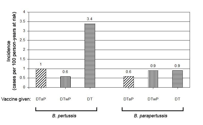
Incidence of B. pertussis and B. parapertussis in German study population, by vaccine group and species infecting. DTaP – diphtheria-tetanus-acellular pertussis vaccine; DTwP – diphtheria-tetanus-whole-cell pertussis vaccine; DT – diphtheria-tetanus vaccine.

As you can see, while there is a large and statistically significant difference in pertussis incidence (left) between vaccine groups (equating to 72% and 83% vaccine efficacy for aP and wP vaccines, respectively) there is barely any difference between the groups when it comes to the frequency of parapertussis infections (right), and the little difference there is isn’t statistically significant. So the German data paints a picture of the aP vaccine decreasing susceptibility to contracting pertussis, but not affecting susceptibility to parapertussis infection.

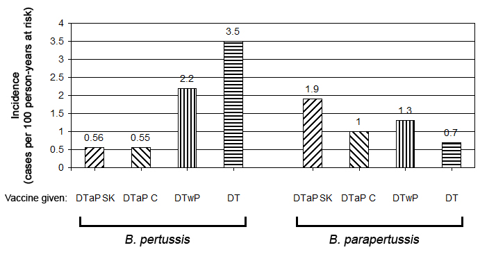
Next is the Italian data. The pertussis-specific data is taken from A controlled trial of two acellular vaccines and one whole-cell vaccine against pertussis. Progetto Pertosse Working Group. The parapertussis-specific data is taken from a follow-up paper by the same authors, in which they analysed the parapertussis data from the same trial, Bordetella parapertussis infection in children: epidemiology, clinical symptoms, and molecular characteristics of isolates

Incidence of B. pertussis and B. parapertussis in Italian study population, by vaccine group. DTaP SB – Smithkline-Beecham DTaP vaccine; DTaP C – Connaught Laboratories DTaP vaccine.

Again, all of the pertussis-immunised groups show a statistically significant difference in pertussis incidence when compared to the control group. However, there are no statistically significant differences in the rate of parapertussis between vaccine groups.

“Now hang on” you might say, “the difference in pertussis incidence between the DTwP and DT groups is about 1.2 cases/100py, and that’s statistically significant, so how is it that the 1.3 cases/100py differnce in parapertussis incidence between the DTaP SK and DT groups isn’t significant?”

Good question. So the calculation for statistical significance isn’t just based on the relative differences between the groups, it’s also based on the strength of the data. This can be factored in in a few ways, such as taking the length of surveillance or number of cases into account (for example, one result based on comparing three cases to three cases may not reach statistical significance, but calculating the same frequency after comparing hundreds to hundreds may). In this case, the pertussis data is based on a total of 288 cases, while the parapertussis is based on a total of 76 cases. I hope that answers your question.

As for the Swedish trial, the collected parapertussis data is not discussed in the study. As with the Italian data, it was discussed in a follow-up paper. However, in this paper they seem more interested in discussing the data they collected on two non-Bordetella organisms that can cause whooping cough. They don’t discuss parapertussis that much, but do say:

B. parapertussis infections fulfilling the clinical WHO criteria
for pertussis were evenly distributed between the vaccine
arms.

I did try to extract data to present graphically as above, but the lack of focus of parapertussis in this article made specifics unclear and my attempt impossible.

So, did the biggest prospective trials of aP vaccination performed in human history find any evidence that susceptibility to B. parapertussis is influenced by receipt of an aP vaccine?

No.

Okay.

Well, I did mention another theoretical outcome of impaired bacterial clearance: worse/longer lasting symptoms.

So: Are acellular pertussis vaccine recipients more susceptible to longer/more serious parapertussis infections?

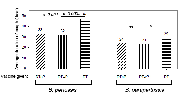
While the other two studies were not as helpful, the German study authors were thoughtful enough to include the breakdown of their pertussis data , with regard to severity of infection.

Average duration of coughing illness by vaccine group and pertussis vs. parapertussis infection. Abbreviations are as explained above. Source

As you can see, there is a statistically significant influence from aP immunisation on the duration of coughing illness caused by B. pertussis. This is consistent with later research showing aP immunisation alleviates disease in cases of pertussis in the immunised. Looking to parapertussis cases we see there is no significant difference in duration of symptoms between the control and pertussis vaccine groups.

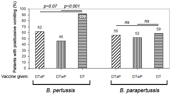
What about severity of disease? One way to measure that is to look at the proportion of patients that have more severe symptoms on infection.

The percentage of patients to experience posttussive vomiting, by vaccine group and causative organism. Source

Posttussive vomiting refers to the act of throwing up following a coughing fit. As you can tell from the ‘DT’ pertussis case data in the above figure, this is common in the unimmunised who catch pertussis, but can be alleviated by immunisation. As for parapertussis cases, there was no statistically significant difference in the proportion of patients to experience posttussive vomiting, regardless of which vaccine group they were randomised to.

Unfortunately, the Italian group did not analyse their parapertussis data by vaccine group. However, they did explore the duration of various symptoms, and how aP immunisation alleviated those caused by B. pertussis. Click here to see the summary. This figure gives a pretty good idea of just how bad pertussis disease can be, and shows how the severity of symptoms is reduced in the vaccinated that do catch the disease.

So, did the largest available prospective aP vaccine trials uncover any evidence to support the hypothesis that the whooping cough shot increases length/severity of symptoms of either kind of whooping cough?

No.

So while it is not ethically possible to test whether the phenomenon (of impaired clearance of parapertussis in the aP immunised) observed in mice also occurs in humans, we can conclude that none of the potential negative manifestations thereof can be seen in human recipients of the aP vaccine in the biggest prospective trials to date.

So never fear – immunising yourself of your child against whooping cough will not raise the chances of catching either cause of whooping cough.

Posted in acellular pertussis, Acellular pertussis vaccine, Bordetella parapertussis, Bordetella pertussis, Pertussis, Pertussis immunisation, Pertussis vaccination, Vaccination, whooping cough immunisation, whooping cough vaccine | 5 Comments